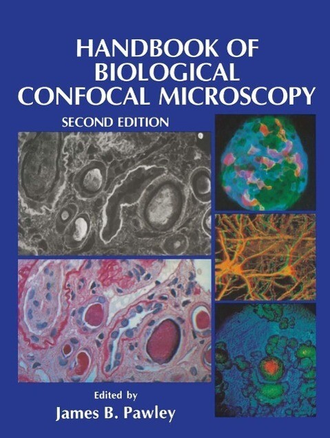
This third edition of a classic text in biological microscopy includes detailed descriptions and in-depth comparisons of parts of the microscope itself, digital aspects of data acquisition and properties of fluorescent dyes, the techniques of 3D specimen preparation and the fundamental limitations, and practical complexities of quantitative confocal fluorescence imaging. Coverage includes practical multiphoton, photodamage and phototoxicity, 3D FRET, 3D microscopy correlated with micro-MNR, CARS, second and third harmonic signals, ion imaging in 3D, scanning RAMAN, plant specimens, practical 3D microscopy and correlated optical tomography.
Inhaltsverzeichnis
1: Foundations of Confocal Scanned Imaging in Light Microscopy. - 2: Fundamental Limits in Confocal Microscopy. - 3: Quantitative Fluorescence Confocal Laser Scanning Microscopy (CLSM). - 4: The Pixilated Image. - 5: Laser Sources for Confocal Microscopy. - 6: Non-Laser Light Sources. - 7: Objective Lenses for Confocal Microscopy. - 8: The Specimen Illumination Path and its Effect on Image Quality. - 9: The Intermediate Optical System of Laser-Scanning Confocal Microscopes. - 10: Intermediate Optics in Nipkow Disk Microscopes. - 11: The Role of the Pinhole in Confocal Imaging System. - 12: Photon Detectors for Confocal Microscopy. - 13: The Collection, Processing, and Display of Digital Three-Dimensional Images of Biological Specimens. - 14: Visualization Systems for Multidimensional CLSM Images. - 15: Mapping and Measuring Surfaces Using Reflection Confocal Microscopy. - 16: Fluorophores for Confocal Microscopy. - 17: Image Contrast in Confocal Light Microscopy. - 18: Guiding Principles of Specimen Preservation for Confocal Fluorescence Microscopy. - 19: Confocal Microscopy of Living Cells. - 20: Lens Aberrations in Confocal Fluorescence Microscopy. - 21: Real-Time Stereo (3D) Confocal Microscopy. - 22: Signal-To-Noise in Confocal Microscopes. - 23: Comparison of Wide-Field/Deconvolution and Confocal Microscopy for 3D Imaging. - 24: Light Microscopic Images Reconstructed by Maximum Likelihood Deconvolution. - 25: Direct View Confocal Imaging Systems Using a Slit Aperture. - 26: Two New High-Resolution Confocal Fluorescence Microscopies (4Pi, Theta) with One- and Two-Photon Excitation. - 27: Optical Considerations at Ultraviolet Wavelengths in Confocal Microscopy. - 28: Two-Photon Molecular Excitation in Laser-Scanning Microscopy. - 29: Video-Rate Confocal Microscopy. - 30: Confocal Microscopy withTransmitted Light. - 31: Fluorescence Lifetime Imaging, a New Tool in Confocal Microscopy. - 32: Imaging Immunogold Labels with Confocal Microscopy. - 33: Fiberoptics in Confocal Microscopy. - 34: Comparison of Various Optical Sectioning Methods. - 35: Mass Storage and Hard Copy. - 36: Tutorial on Practical Confocal Microscopy and Use of the Confocal Test Specimen. - 37: Bibliography of Confocal Microscopes. - Appendix 1: Optical Units. - W. B. Amos. - Radial units. - Axial units. - Appendix 2: Light Paths of Current Commercial Confocal Light Microscopes for Biology. - James Pawley.


