Sofort lieferbar (Download)
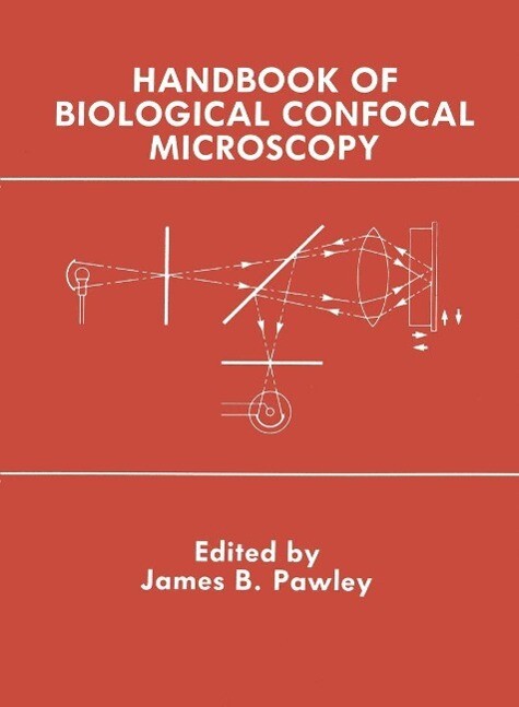
In 1987 the Electron Microscopy Society of America (EMSA) going to drive important scientific discoveries across wide areas under the leadership of J. P. Revel (Cal Tech) initiated a major of physiology, cellular biology and neurobiology. They had been program to present a discussion of recent advances in light looking for a forum in which they could advance the state of microscopy as part of the annual meeting. The result was three the art of confocal microscopy, alert manufacturers to the lim special LM sessions at the Milwaukee meeting in August 1988: itations of current instruments, and catalyze progress toward The LM Forum, organized by me, and Symposia on Confocal new directions in confocal instrument development. LM, organized by G. Schatten (Madison), and on Integrated These goals were so close to those of the EMSA project that Acoustic/LM/EM organized by C. Rieder (Albany). In addition, the two groups decided to join forces with EMSA to provide there was an optical micro-analysis session emphasizing Raman the organization and the venue for a Confocal Workshop and techniques, organized by the Microbeam Analysis Society, for NSF to provide the financial support for the speakers expenses a total of 40 invited and 30 contributed papers on optical tech and for the publication of extended abstracts.
Inhaltsverzeichnis
1: Foundations of Confocal Scanned Imaging in Light Microscopy. - Light microscopy. - Lateral resolution. - Axial resolution. - Depth of field. - Confocal imaging. - Impact of video. - The Nipkow disk. - Electron-beam scanning TV. - Impact of modern video. - Lasers and microscopy Holography. - Laser illumination. - Laser-illuminated confocal microscopes. - Laser scanning confocal microscope. - Is laser scanning confocal microscopy a cure-all? . - Speed of image or data acquisition. - Depth of field in phase dependent imaging. - Some other optical and mechanical factors affecting confocal microscopy. - Lens aberration. - Unintentional beam deviation. - Note added in proof. - Acknowledgment. - References. - 2: Fundamental Limits in Confocal Microscopy. - What limits? . - Counting statistics. - Source brightness. - Specimen response. - A typical problem. - Practical photon efficiency. - Losses in the optical system. - Objectives. - Mirrors. - Pinhole. - Is the confocal pinhole a good thing ? . - Features of the confocal pinhole. - Detection and measurement losses. - The detector. - The PMT. - Solid-state photon detectors. - Digitization. - Evaluating photon efficiency. - Resolution: how much is enough? . - Can resolution be too high? . - Limitations imposed by spatial and temporal quantization. - Aliasing. - Pixel shape. - Blind spots. - Practical considerations relating resolution to distortion. - Summary. - Acknowledgements. - References. - 3: Quantitative Fluorescence Imaging with Laser Scanning Confocal Microscopy. - The promise of scanning confocal fluorescence microscopy. - Optical transfer efficiency. - Methods of measurement of optical transfer efficiencies. - Confocal spatial filtering for depth of field compromises optical transfer efficiency. - Optical aberrations in fluorescence LSCM. - Chromatic aberrations influorescence LSCM measurements. - Photodynamic effects. - Theory. - Population rate equations. - Experiment. - Fluorescence photobleaching recovery with LSCM. - Conclusion. - Acknowledgments. - References. - 4: The Pixelated Image. - Pixelation. - Optical resolution: the resel. - Pixel. - Gray level. - In between. - Matching image spatial characteristics. - The Nyquist theorem. - Hyper-resolution: oversampling. - The resel/pixel ratio. - Diagonal dropout. - Pixel shape distortion. - Aliasing. - The mechanics of pixelation. - Matching image intensity characteristics. - Gray scale. - Detection. - Display. - Color displays. - Caveats. - Spectral variation of detectors. - Automatic gain control. - Strategy for magnification and resolution. - Acknowledgements. - References. - 5: Laser Sources for Confocal Microscopy. - Laser power requirements. - The basic laser. - Principle of operation. - Laser modes: longitudinal and transversal. - Polarization. - Coherent properties of laser light. - Temporal coherence. - Coherence length. - Spatial coherence. - Coherence surface. - Coherence volume. - Pumping power requirements. - Heat removal. - Other installation requirements. - Types of lasers. - Continuous wave (cw) lasers. - Gas lasers. - Argon-ion. - Krypton. - Helium-neon. - Helium-cadmium. - Dye lasers. - Solid state lasers. - Semiconductor or diode injection lasers. - Diode-pumped lasers. - Tunable solid state laser. - Pulsed lasers. - Nitrogen lasers. - Excimer lasers. - Metal vapor lasers. - Q-switched lasers. - Trends in time-resolved spectroscopy applied to microscopy. - Wavelength expansion through non-linear techniques. - Spatial beam characteristics. - Intensity fluctuations of cw lasers. - Maintenance. - Maintenance of active laser media. - Laser tubes. - Gases. - Dyes. - Laser rods. - Maintenance of pumping media. - Maintenance of the opticalresonator. - Maintenance of other system components. - Cooling water. - External optics. - Safety precautions. - Curtains. - Screens. - Beam stops. - Acknowledgement. - References. - 6: Non-Laser Illumination for Confocal Microscopy. - Why use non-laser sources? . - Wavelength. - Coherence. - Which types of confocal microscope can use nonlaser sources? . - Characteristics of non-laser light sources. - Wavelengths available. - Source radiance. - Source stability. - Source coherence. - Source distribution. - Collecting the light and relaying it to specimen. - Illumination of the specimen: a basic part of microscopy. - Tandem scanning: basic description. - Single-sided disk scanning: basic description. - How do you uniformly illuminate both the objective back focal plane and the intermediate image plane? . - Scrambling and filtering the light. - Measuring what comes through the illumination system. - Exposure time and source brightness. - Stationary specimens. - What if the specimen is moving or changing? . - Incoherent laser light sources for confocal microscopy. - References. - 7: Objective Lenses for Confocal Microscopy. - Abstract. - Aberrations of refractive systems. - Defocusing. - Monochromatic aberrations. - Spherical aberrations. - Coma. - Astigmatism. - Flatness of field. - Distortion. - Chromatic aberrations. - Longitudinal chromatic aberration. - Lateral chromatic aberration (LCA) or chromatic magnification difference. - Finite versus infinity optics. - Optical materials. - Anti-reflection coatings. - Conclusion. - 8: Size and Shape of the Confocal Spot: Control and Relation to 3d Imaging And Image Processing. - Abstract. - Pinholes and optical probe formation. - Practical use of variable pinholes. - Experimental axial confocal response. - Comments and conclusions. - References. - 9: The Intermediate Optical System of Laser-Scanning Confocal Microscopes. - Design principles of confocal systems. - Overview. - Microscope objectives. - Position of the pivot point. - Position of the detector pinhole. - Practical requirements. - Illumination. - Detection. - Distortion. - Evaluation of illumination/detection systems. - Influence of optical elements on the properties of light. - Errors caused by optical elements. - Evaluation of optical arrangements. - Evaluation of scanner arrangements. - Disk scanners. - Object scanners. - Attachment to microscopes. - Merit functions. - Requirements for multi-tluorescence experiments. - Special optical elements. - Multi-mode optical glass fibers. - Single-mode polarization-preserving glass fibers. - Polarizing elements. - Mechanical scanners. - Acousto-optical scanners. - Conclusions. - Acknowledgements. - References. - 10: Intermediate Optics in Nipkow Disk Microscopes. - The tandem scanning reflected light microscope (TSRLM). - The real-time scanning optical microscope (RSOM). - Images of the eye. - Pinhole size. - Pinhole spacing. - Illumination efficiency and reflection from the disk. - Internal reflections. - Acknowledgements. - References. - 11: The Role of the Pinhole in Confocal Imaging Systems. - The optical sectioning property. - The optical sectioning property with a finite-sized circular detector and coherent light. - Lateral resolution as a function of effective detector size. - The role of aberrations. - Images with a finite-sized detector 118 Extended-focus and auto-focus imaging with a finite-sized detector. - Height imaging with a finite-sized pinhole. - Alternative detector geometries. - Noise. - Fluorescence imaging. - Conclusions. - References. - 12: Photon Detectors for Confocal Microscopy. - The quantal nature of light. - Interaction of photons with materials. - Photoconductivity. - Photovoltaic. - Charge coupled devices. - Photoemissive. - Image dissector l. - Micro channel plate. - Noise internal to detectors. - Statistics of photon flux and detectors. - Representing the pixel value. - Conversion techniques. - Assessment of devices. - Point detection optimization. - Field detection optimization. - Detectors present and future. - References. - 13: Manipulation, Display, and Analysis of three-Dimensional Biological Images. - Storage of three-dimensional image data. - Image enhancement. - Linear filters. - Median filters. - Local contrast enhancement. - Gradient method. - Processing methods for displaying 3D data. - Stereo images. - 3D rotations. - Rotated projections. - Pixar displays. - Contour surface representation. - Graphic system for 3D image display and analysis. - Details of PRISM s design and implementation. - The window system. - Digital movies. - Choice of display hardware. - Model building in PRISM. - Model building. - Superimposing the model on a background image. - Future development and discussion. - Acknowledgements. - References. - 14: Three-Dimensional Imaging on Confocal and Wide-Field Microscopes. - Signal to noise ratio and resolution. - Signal strength, photo-damage and photo-bleaching. - Optical transfer function, resolution and noise. - Three-dimensional image restoration and confocal microscopy: a comparison. - Image restoration methodology. - Requirements. - Results. - Multiple detector confocal miscroscopes. - Conclusions and recommendations. - Computer graphics 3D visualization. - Display of 2D slices of 3D data. - Volume displays. - Surface model displays. - 3D perception from 2D displays. - User interaction and analysis. - Automated image analysis: feature extraction and computer vision. - Thresholding. - Human interaction and partial automation. - Fully automated analysis. -Computer hardware considerations. - Image acquisition. - Image restoration. - Image analysis and display. - Archival storage. - Magnetic disks. - Cartridge magnetic tape. - Reel magnetic tape. - Optical disk. - Networking. - Conclusion. - References. - 15: Direct Recording of Stereoscopic Pairs Obtained Directly from Disk Scanning Confocal Light Microscopes. - Summary. - Use of a confocal microscope to reduce the depth of field. - Optical sectioning in the TSRLM. - Direct photographic recording of the stereo-pair. - Means for stereo imaging of a layer inside a bulk. - Fixed tilt angle difference: hand-operated device. - DC micromotor-controlled stage. - Piezo-electric control of the lens. - Top or bottom overlap? . - Stereopairs generated from one through-focus pass. - Topographic mapping. - Color coding without a computer. - Depth limitation. - Particle counting. - Geometric properties of the stereo images. - Discussion: TSRLM or LSCM? . - Acknowledgements. - References. - 16: Fluorophores for Confocal Microscopy: Photophysics and Photochemistry. - Photophysical problems related to high intensity excitation. - Singlet state saturation. - Triplet state saturation. - Contaminating background signals. - Rayleigh and Raman scattering. - A utofluorescence from endogenous fluorophores. - What is the optimal intensity? . - Photodestruction of fluorophores and biological specimens. - Dependency on intensity or its time integral? . - Theory. - Experiment. - Protective agents. - Strategies for signal optimization in the face of photobleaching. - Light collection efficiency. - Spatial resolution. - Fluorophore concentration. - Choice of fluorophore. - Fluorescent indicators for dynamic intracellular parameters. - Membrane potentials. - Ion concentrations. - Wavelength ratioing. - pH indicators. - Ca2+ indicators. - Other forms of ratioing. - Future developments? . - Acknowledgments. - References. - 17: Image Contrast in Confocal Light Microscopy. - Sources of contrast. - Confocal microscopy in back scattered mode. - Signal formation. - Backscattered light contrast on stained specimens. - Reflection contrast on non-biological specimens. - Backscatter contrast on living specimens. - The effect of overlying structures. - Absorption contrast. - Artificial contrast. - Transmitted confocal image. - Confocal microscopy in epi-fluorescent mode. - Countermeasures. - Acknowledgement. - References. - 18: Guiding Principles of Specimen Preservation for Confocal Fluorescence Microscopy. - Critical evaluation of fixation and mounting methods. - Theoretical considerations. - The use of the cell height to evaluate the fixation method. - The use of cell height to evaluate mounting media. - Well defined structures can be used to evaluate fixation methods. - Comparison of in vivo labeled cell organelles with immunolabeled cell organelles. - Fixation methods. - Glutaraldehyde fixation. - Stock solutions. - Preparation of stock solutions. - Fixation protocol. - The pH shift/paraformaldehyde fixation. - Stock solutions. - Preparation of the stock solutions. - Fixation protocol. - Immunofluorescence staining. - Mounting the specimen. - General notes. - Labeling samples with two or more probes. - Ramifications of techniques to preserve the specimens. - Conclusion. - Acknowledgements. - References. - 19: A Comparison of Various Optical Sectioning Methods: The Scanning Slit Confocal Microscope. - Non-Confocal Optical Sectioning. - Confocal Optical Sectioning. - Scanning Mifror/Slit Microscope. - Optical Sectioning: Experimental and Theoretical. - Scanning Mirror/Slit System. - Confocal Pinhole System. - Theoretical Comparison of Slit and Pinhole Systems. - A Possible Improvement in Pinhole Confocal Systems. - A Possible Improvement in the Slit Scanning System. - Examples of Images Obtained with the Divided Aperture Scanning Slit System. - Summary Comparison of Slit and Pinhole Confocal Svstems. - Slit system. - Inherent advantages. - Practical advantages of divided aperture system as described. - Single pinhole confocal systems. - Inherent advantages. - References. - Bibliography on Confocal Microscopy.
Produktdetails
Erscheinungsdatum
06. Dezember 2012
Sprache
englisch
Seitenanzahl
232
Dateigröße
46,59 MB
Herausgegeben von
James Pawley
Verlag/Hersteller
Kopierschutz
mit Wasserzeichen versehen
Produktart
EBOOK
Dateiformat
PDF
ISBN
9781461571339
Entdecken Sie mehr
Bewertungen
0 Bewertungen
Es wurden noch keine Bewertungen abgegeben. Schreiben Sie die erste Bewertung zu "Handbook of Biological Confocal Microscopy" und helfen Sie damit anderen bei der Kaufentscheidung.


